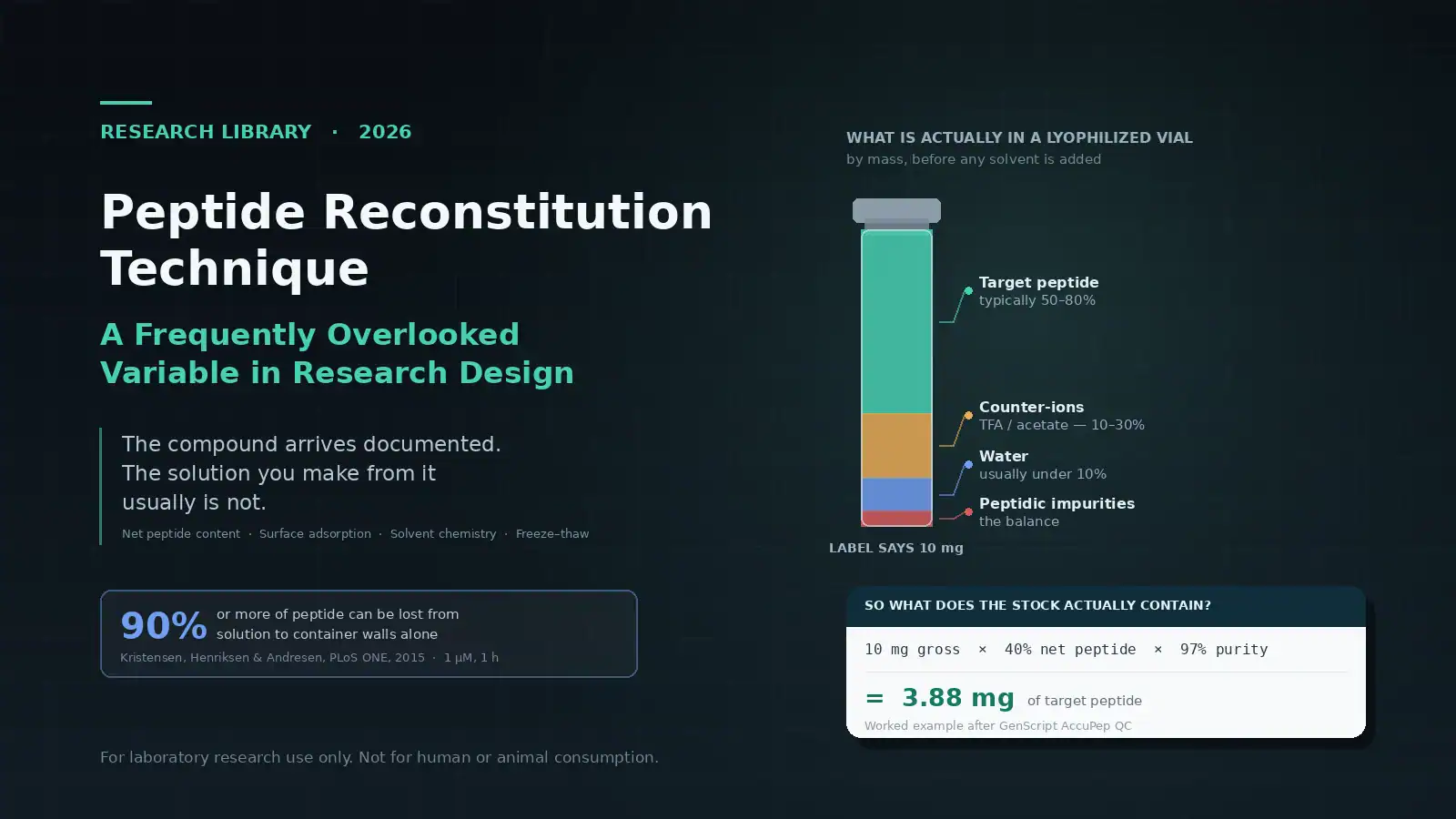

A vial labeled 10 mg is not a vial containing 10 mg of peptide — and no amount of careful pipetting corrects for the difference.
Every document that accompanies a research peptide describes a solid. The certificate of analysis characterises a powder. The mass spectrum confirms the identity of a powder. The chromatogram reports the purity of a powder.
Then solvent goes into the vial, and all of that documentation stops describing the thing in your hands.
What you have afterwards is a solution, and a solution has properties the powder did not: a concentration that depends on how much of the powder was actually peptide, a recovery that depends on what the container is made of, a stability that depends on pH and temperature history, and a composition that changes every time it is frozen and thawed. None of those properties appear on the COA, because none of them existed when the COA was written.
Reconstitution is the handoff point between a characterised material and an uncharacterised one. It is also, in most protocols, the step described in the fewest words.
Peptides Exchange sells research peptides, which is a reason to read what follows with appropriate scepticism. The honest version of this topic is not flattering to suppliers, this one included — the largest error source described below is one the supplier controls and the customer usually cannot see. Where that is the case, this article says so.
New to the category? What Are Research Peptides? is the better starting point. For the documentation side, see How to Read a Peptide Certificate of Analysis.
A note on scope
This article covers the preparation of stock solutions for in-vitro laboratory work — cell culture, binding assays, analytical standards, stability studies. It deals with solvent chemistry, recovery, and documentation.
It contains no reconstitution volumes, no concentration calculators, and no vial arithmetic, and it does not discuss diluents intended for injectable preparations. That omission is deliberate and is explained in the regulatory section at the end.
The error no pipette can correct: net peptide content
Start with the largest source of concentration error, because it dwarfs the others and because most protocols never account for it.
A lyophilized peptide vial does not contain only peptide. By mass, a typical vial breaks down roughly as follows:
| Component | Typical share of mass |
|---|---|
| Target peptide | 50–80% |
| Counter-ions (TFA, acetate, hydrochloride) | 10–30% |
| Water | usually under 10%, higher for hydrophilic peptides |
| Peptidic impurities | the balance |
Those ranges come from GenScript’s technical material on peptide sample composition. The fraction that is actually target peptide is called net peptide content, and Bachem defines it as “the percentage of peptides relative to nonpeptidic material, mostly counter-ions and moisture.”
Why purity does not tell you this
Purity and net peptide content measure different things, and conflating them is the most common error in this whole subject.
RP-HPLC purity is an area percentage: the target peak as a share of all UV-absorbing peaks detected. It is a statement about the relative abundance of the species the detector can see.
Trifluoroacetate does not absorb meaningfully at the wavelengths used, so it does not appear as a peak. Neither does water. A peptide can therefore be 99% pure by HPLC and still be 60% peptide by mass — the chromatogram is not wrong, it is answering a different question. Bachem states the point plainly: net peptide content “includes peptidic contaminants” and so is not equivalent to purity, and peptides rich in basic residues “can have a low NPC even when very pure.”
This is the same identity-versus-purity-versus-content distinction covered in HPLC vs. LC-MS Peptide Testing — three questions, three methods, no substitutions.
What that does to a stock solution
Work the arithmetic through. A vial labeled 10 mg gross, with net peptide content of 40% determined by amino acid analysis and HPLC purity of 97%:
> 10 mg × 40% = 4.0 mg of peptide
> 4.0 mg × 97% = 3.88 mg of target peptide
Dissolve that vial and treat it as 10 mg, and every concentration downstream is overstated by a factor of about 2.6. Every IC₅₀, every EC₅₀, every dose–response curve shifts. The experiment is internally consistent and externally meaningless, and nothing about the technique used to dissolve it is at fault.
Bachem gives the general relationship as: absolute peptide quantity = quantity of lyophilizate × purity × net peptide content.
Why basic peptides are worst affected
Counter-ion load is not random. Peptides are supplied as TFA salts by default, because TFA is the standard ion-pairing agent in preparative RP-HPLC. TFA associates with basic sites — arginine, lysine, histidine, and the free N-terminus. The more basic residues a sequence carries, the more counter-ion mass rides along with it.
So the peptides with the lowest net peptide content tend to be exactly the arginine- and lysine-rich sequences, which includes a substantial share of the research market. Bachem notes that such peptides “can carry a considerable number of counter-ions,” and that TFA “cannot be fully removed” without a separate ion-exchange step.
Water compounds the problem. Lyophilizates hold varying amounts of non-covalently bound water, and peptides — hydrophilic ones especially — are hygroscopic. A vial opened at room temperature in a humid room gains mass that is not peptide. Bachem’s handling guidance is to let the sealed vial reach ambient temperature in a desiccator before opening, weigh quickly, and reseal tightly.
What to do about it
There are only two honest options.
Obtain the net peptide content. It is determined by amino acid analysis — the basis of USP General Chapter ⟨1052⟩ — or by elemental nitrogen (CHN) analysis, which works because counter-ions and water contain no nitrogen. A supplier that has run it can provide the number. One that has not, cannot.
Or state your concentrations as nominal. If the net peptide content is unknown, then the stock concentration is unknown, and the correct way to report it in a methods section is as a nominal concentration based on labeled mass, with the limitation stated. That is an honest record. Treating a nominal figure as a measured one is not.
Net peptide content is item eight on the Research Peptide Supplier Evaluation Checklist, and it is one of the checks that most certificates in this market fail — including, as that article notes, some in our own catalogue.
Solvent selection is chemistry, not preference
Once you know what is in the vial, the next question is what will dissolve it. This is determined by the sequence, not by habit.
The net-charge method
Sigma-Aldrich’s peptide handling guidelines set out the standard approach. Calculate net charge by assigning −1 to each acidic residue (Asp, Glu, and the C-terminal carboxyl), +1 to each basic residue (Lys, Arg, and the free N-terminal amine), and +1 to each histidine at pH below 6. Then:
| Net charge | Approach |
|---|---|
| Negative (acidic peptide) | Dissolve in a small volume of a mildly basic medium — 0.1 M ammonium bicarbonate, or dilute aqueous ammonia — then dilute toward neutral |
| Positive (basic peptide) | Dissolve in a small volume of a mildly acidic medium such as dilute acetic acid, then dilute |
| Zero, with charged residues >25% of the sequence | Treat as acidic |
| Zero, with charged residues 10–25% | Organic solvent first, then dilute |
| Charged residues <10% (hydrophobic) | Organic solvent — acetonitrile, DMSO or DMF; chaotropes such as urea or guanidine hydrochloride for gelling sequences |
Bachem’s guidance is consistent: dissolve in a small amount of the appropriate solvent first, then dilute into the working buffer, noting that phosphate-buffered saline near pH 7.0–7.4 is the safest diluent where concentrations of 1 mg/mL or lower are sufficient.
The sequencing matters. Adding the working buffer directly to a stubborn peptide and then trying to rescue it tends to produce a suspension that looks dissolved. Dissolving in the minimum volume of the right solvent first, then diluting, is the step most often skipped.
Test a portion first
Sigma’s guidance is explicit, and it is worth quoting: “it is necessary to test a portion of the peptide first before dissolving the entire peptide sample.”
This is the cheapest insurance in the whole procedure, and the one most often skipped under time pressure. A solvent choice that turns out to be wrong after the entire vial is committed cannot be undone.
Dissolved or suspended?
A solution that gels, looks cloudy, or shows particulates is a suspension, not a solution. Sigma’s guidance is to sonicate briefly before escalating to a stronger solvent, since sonication breaks the solid into smaller particles and can confirm whether the material is genuinely insoluble. Bachem adds that dissolution may take hours, that brief water-bath sonication can help, and that excessive warming should be avoided.
The practical risk is drawing an aliquot from a suspension. The concentration of that aliquot is whatever the local particle distribution happened to be, and it will not match the next one.
The DMSO ceiling
DMSO solves solubility problems and creates biological ones. It is not inert, and in cell-based work the solvent concentration is itself an experimental variable.
Santos, Shimabuko and Sipert tested DMSO at 0.1%, 0.5%, 1%, 5% and 10% against apical papilla cells over 24 hours to 7 days, scoring cytotoxicity by the ISO 10993-5 criterion of a greater-than-30% reduction in viability (Brazilian Dental Journal, 2024). At 0.1% there was no significant reduction at any time point. At 0.5% there was none except at 72 hours, and not past the cytotoxicity threshold. At 5% and above, DMSO was cytotoxic at every time point. The authors conclude that DMSO “ideally should not exceed 0.5% of the total medium volume,” and recommend a DMSO-matched control group in pharmacological studies.
That matched control is the part that gets dropped. If the treatment wells receive 0.4% DMSO and the control wells receive none, the experiment compares peptide-plus-solvent against nothing, and the solvent’s own effect is inside the result. The vehicle control must carry the same final solvent concentration as the highest treatment condition — and the final concentration, not the stock concentration, is what matters.
Two further constraints: Bachem notes that high organic solvent concentrations are incompatible with cells generally, and Sigma notes that peptides containing cysteine or methionine are unstable in DMSO. For a free-thiol peptide, DMSO is not a neutral choice of solvent; it is an oxidising environment.
Oxidation-prone sequences
Peptides containing Cys, Met or Trp need more care at the solvent step than the rest.
Cysteine thiols oxidise rapidly above pH 7, forming disulfides. Bachem recommends dissolving free-cysteine peptides in carefully degassed acidic buffers, and using oxygen-free water or a reducing agent such as DTT or TCEP where appropriate. Disulfide formation is reversible with those reagents.
Methionine oxidation is not. Met oxidises to the sulfoxide and then the sulfone, and Sigma describes these products as “almost impossible to reverse.” A methionine-containing peptide handled carelessly at reconstitution is permanently altered, and the alteration is a separate molecular species with its own behaviour.
This is not an abstract concern for specific sequences. MOTS-c, for example, contains methionine at three positions, which is why the WADA detection method validates its oxidation products as separate analytes — covered in What Is MOTS-c?.
The peptide that disappears: surface adsorption
This is the variable researchers most often discover by accident, after a dose–response curve comes back flat.
Peptides adsorb to container walls. The effect is large, it is worst at exactly the low concentrations that matter in assay work, and it is not fixed by good technique.
How large
Kristensen, Henriksen and Andresen measured recovery of three cationic peptides — mastoparan X, melittin and magainin 2 — from borosilicate glass, standard polypropylene, and Protein LoBind tubes (PLoS ONE, 2015, 10(5):e0122419). Their headline finding is that at typical working concentrations, “90% or more of the peptides might be lost from solution.”
The specifics:
- In 220 µL of 1 µM solution incubated for one hour, glass and polypropylene recovered only 10–20% of the peptide.
- Across successive transfers of 5 µM solutions between glass or polypropylene containers, recovery approached zero after four transfers.
- In 2 mL of 2 µM solution, quartz cuvettes recovered 50% or less for all three peptides.
Protein LoBind tubes lost less than standard glass and polypropylene, but did not eliminate the effect.
A second study makes the concentration dependence concrete. Weikart and colleagues compared recovery of peptides and proteins on standard polypropylene, a commercial low-bind plate, and plasma-treated ultra-low-binding microplates (Future Science OA, 2019, 5(2):FSO367). At 1.5 nM, EGF recovery was 20% on standard polypropylene versus 100% on the low-binding surfaces. Glucagon recovered at roughly 5–10% across the range on both standard and low-bind plates, and 71% on the ultra-low-binding plate. Insulin recovery on the commercial low-bind plate fell from 95% at 12.5 nM to 74% at 1.5 nM — the same plate, the same peptide, a recovery difference created purely by dilution.
That study was conducted by the manufacturer of the plates tested, which is worth weighing. The direction of the effect, though, is consistent with the independent PLOS ONE work.
What actually helps
From the Kristensen data:
Low-binding consumables help but do not solve it. They reduce loss; they do not abolish it.
Higher volume-to-surface ratio helps. Less relative loss occurs in larger volumes, because the wall area per unit volume is smaller.
Higher concentration helps, because the walls saturate. This is why the problem is invisible when you prepare a concentrated stock and appears only at the working dilution.
Ionic strength does not help. Adjusting NaCl did not meaningfully reduce adsorption.
Pre-saturating the container is unreliable. Adsorption was partly reversible in these experiments, which means walls loaded with peptide can release it back into a later solution. A pre-rinsed tube is not a clean tube; it is a tube with an unknown amount of peptide ready to come off.
The practical consequence is that adsorption should be treated as a property of the assay system and controlled for, not technique-corrected away. If the working concentration is in the nanomolar range, the concentration in the well is not the concentration you calculated, and the gap between them depends on the plastic, the volume, the incubation time and the number of transfers.
Freeze–thaw is a chemical event, not a pause
“Store frozen in aliquots” is standard advice. The reason it is standard is less widely known, and the mechanism is more aggressive than most protocols assume.
The pH shift
Thorat, Munjal, Geders and Suryanarayanan examined freezing-induced aggregation and traced it to a pH shift caused by selective buffer crystallization (Journal of Controlled Release, 2020, 323:591–599).
In 100 mM sodium phosphate buffer, cooling from 20 °C to −25 °C dropped the pH by 3.1 units for BSA solutions and 2.7 units for β-galactosidase. The cause is that disodium hydrogen phosphate crystallizes out as the dodecahydrate, confirmed by low-temperature X-ray diffraction. Removing the basic component from the remaining liquid phase drives the pH of that phase down sharply. Micro-flow imaging showed a marked rise in particle counts after freeze–thaw cycling.
A three-unit pH swing is not a storage condition. It is an excursion well outside the range in which most peptides are stable, happening inside the tube, every cycle.
The authors found two mitigations: adding a non-crystallizing solute (cellobiose) limited the shift to about 1.0 unit with no aggregation observed, and simply lowering the buffer concentration to 10 mM prevented aggregation. The second is the more useful finding for routine work — a concentrated phosphate buffer is a worse freezing medium than a dilute one, which is the opposite of the intuition that more buffer means more buffering.
Why one thaw per aliquot
Sigma’s guidance is direct: “Avoid repeated freeze-thaw cycles, as this can degrade the peptides,” with individual aliquots frozen so that each is thawed only once. Bachem’s position is firmer still — peptides should not be stored in solution at all, even sterile and oxygen-free, because they “may slowly chemically degrade,” and frozen solutions should be regarded as keeping only “for a few weeks.”
Both recommend aliquoting before freezing and storing below −15 °C, with sterile buffers around pH 5–6 extending solution life.
Deamidation runs on its own clock
Freezing slows chemistry; it does not stop it, and some reactions proceed in solution at any temperature.
Asparagine and glutamine deamidate through a base-catalysed mechanism that proceeds via a cyclic imide intermediate, and the rate is strongly sequence-dependent — Asn-Gly and Gln-Gly motifs are the classic fast cases. The products include aspartate and iso-aspartate analogues, and Sigma notes that some of these forms may be inactive.
Iso-aspartate formation is the detail worth holding onto. It inserts an extra methylene into the backbone, changing the peptide’s shape without changing its mass by much. A deamidated peptide can therefore look close to correct by some analytical measures while behaving differently in an assay.
Sigma lists Asn, Gln, Cys, Met and Trp as the residues that make a peptide “unstable when stored in solution.” A sequence containing any of them deserves a shorter solution lifetime and a documented one.
Mechanical handling
Three short points, because this is where most existing writing on reconstitution begins and ends.
Dissolution takes time. Bachem notes it may take hours. Escalating to vigorous agitation because a peptide has not dissolved in thirty seconds trades a solubility problem for an aggregation problem.
Sonication should be brief and cool. A short water-bath sonication aids dissolution; prolonged sonication deposits energy as heat, and warming a peptide solution accelerates every degradation pathway described above.
Gentle mixing is not superstition. Vigorous agitation introduces air–liquid interface, and interfacial stress is a well-characterised driver of aggregation and subvisible particle formation in protein and peptide solutions. Swirling and passive dissolution avoid generating that interface. This is also why a solution that has been vortexed hard and then looks slightly hazy should not be assumed to have simply “not finished dissolving.”
Volume: the one error with a standard behind it
Of the four variables in this article, volumetric error is both the smallest and the only one governed by a formal metrological standard. That asymmetry is itself informative.
ISO 8655 covers piston-operated volumetric apparatus. The 2022 revision expanded it to nine parts — terminology and user requirements, pipettes, burettes, dilutors, dispensers, the gravimetric reference procedure, alternative procedures, the photometric reference procedure, and manually operated precision laboratory syringes. Under the reference procedures, a full calibration requires a minimum of ten measurements at each of three volumes — 100%, 50% and 10% of nominal — the so-called 10.3 calibration, using at least two tips per volume. The revision also tied calibration results to the specific tip type used, tightened balance-resolution requirements, and dropped the gravimetric method’s status as the preferred reference (summary of ISO 8655:2022).
The useful comparison: a pipette operating within specification contributes a volumetric error typically in the low single-digit percentages. An unaccounted net peptide content of 40% contributes an error of 150%. Adsorption at nanomolar concentration can contribute 80–90%.
Pipette calibration is worth doing, and it is worth recording. It is not where the concentration went.
What to record
Reconstitution is a documentation problem as much as a technique problem, and the documentation failure is the one that compounds — an unrecorded variable cannot be investigated later, and the experiment cannot be repeated by anyone, including the person who ran it.
This is a recognised problem at the level of the literature. Batista Leite and colleagues, writing for the PRO-MaP initiative, note that “essential details about study methods are often missing from academic research papers in the life sciences,” weakening reproducibility and preventing method reuse (PLOS Biology, 2024, 22(9):e3002835). Their recommendations include writing and sharing step-by-step protocols in versioned repositories, using research resource identifiers to specify reagents unambiguously, and documenting protocols in enough detail for others to reproduce them.
A reconstitution record that satisfies that standard captures:
| Field | Why it matters |
|---|---|
| Compound, supplier, catalogue and lot number | The lot is what ties the solution to a specific COA; net peptide content varies batch to batch |
| Labeled mass and net peptide content, with method | Determines whether your concentration is measured or nominal |
| HPLC purity and the method it came from | Separate figure, separate question |
| Solvent, grade, and any pH adjustment | Reproducibility; also the basis of the vehicle control |
| Final solvent concentration in the assay | What the vehicle control must match |
| Resulting concentration, flagged nominal or measured | The single most important line |
| Container type and material for stock and working dilutions | Determines adsorption behaviour |
| Dissolution method and time, including any sonication | Distinguishes dissolved from suspended |
| Date and time of reconstitution | Starting point for every degradation clock |
| Storage temperature and buffer composition | Freeze-concentration and pH-shift risk |
| Number of freeze–thaw cycles for this aliquot | Rarely recorded, frequently the explanation |
| Appearance at reconstitution and at each use | Haze or particulates invalidate the assumed concentration |
The freeze–thaw count is the field most often missing and the one most often needed. Two aliquots of the same stock, one thawed once and one thawed five times, are not the same reagent, and nothing in the record distinguishes them unless someone wrote it down.
When reconstitution is the explanation
A few patterns point back at the preparation step rather than the biology:
A dose–response curve that is flat at the low end but normal at high concentrations. Consistent with adsorption — the walls take a fixed quantity, which is a large fraction of a small amount and a small fraction of a large one.
Potency that drifts downward across a series of experiments from one stock. Consistent with cumulative freeze–thaw, or with degradation of an Asn/Gln/Met/Cys/Trp-containing sequence in solution.
A result that does not reproduce from a new lot at the same nominal concentration. Consistent with a net peptide content difference between batches. This is the one to check first when a finding fails to replicate after reordering.
An effect that persists in the vehicle control. Solvent, not peptide.
Results that differ between two labs following the same written protocol. Consistent with anything in the table above that the protocol did not specify — most often container type or solvent sequence.
None of these is diagnostic on its own. All of them are cheaper to check than to re-run the biology.
What a supplier should provide
The customer-side variables in this article are controllable. The supplier-side one — net peptide content — is not, and no amount of laboratory care compensates for its absence.
A supplier should be able to provide, per lot: the certificate of analysis with the lot number matching the vial, the RP-HPLC chromatogram with its method, mass spectrometric identity confirmation, and net peptide content with the method used to determine it. The twelve-item version of that list is in the Research Peptide Supplier Evaluation Checklist, and our own certificates are posted at Certificates of Analysis.
Our catalogue is organised by research area at the shop, including metabolic, mitochondrial and cellular energy, regeneration and longevity, and cognitive and neuro categories. Compound-specific handling considerations are covered in the individual guides — What Is BPC-157?, What Is MOTS-c?, and the Cagrilintide Research Guide among them. Multi-component preparations raise their own reconstitution questions, discussed in Best Peptide Blend Suppliers.
All products are supplied for laboratory research use only.
<a id=”why-this-article-contains-no-volumes”></a>
Why this article contains no volumes
A reader who came here looking for “how much liquid goes in the vial” will not find it, and the reason is worth stating directly rather than leaving as a gap.
Under 21 CFR 201.128, the intended use of a product is determined from the objective intent of the people who sell it, inferred from the whole net impression their materials create — not from a disclaimer. A seller’s own words, labelling and surrounding content can establish that a product is intended for human use regardless of what the “research use only” line says.
In its enforcement during 2026, FDA cited specific content patterns as evidence of that intent. The April 8, 2026 round of warning letters, five of which went to sellers marketing products as research-use-only, named reconstitution instructions, reconstitution calculators, dosing guidance, and bacteriostatic water marketed alongside the peptides among the materials establishing intended human use. The August 24, 2026 letters repeated the point, treating a diluent sold next to a peptide with preparation guidance as supplying the means to prepare an injectable product for human administration.
A volume figure or a calculator on this page would serve one purpose, and the regulator has already said which purpose it reads them as serving. Laboratory stock preparation does not require a published volume — the concentration needed depends on the assay, and the researcher calculates it from the mass they actually have. Which, as the first section of this article argues, is the number most people do not have.
This is also why this article does not link or discuss diluent products, and does not discuss preparations for injection. Neither omission is an oversight.
For laboratory research use only. Not for human or animal consumption. Not for diagnostic or therapeutic use. Not intended to diagnose, treat, cure or prevent any disease. Peptides Exchange is a chemical supplier and is neither a 503A compounding pharmacy nor a 503B outsourcing facility.
Frequently asked questions
Does a certificate of analysis tell me how much peptide is in the vial?
Not unless it reports net peptide content specifically. Purity is an area percentage from the chromatogram and says nothing about counter-ion or water mass. A COA showing 99% purity is compatible with a vial that is 60% peptide by mass.
What is net peptide content and how is it measured?
It is the fraction of the lyophilizate’s mass that is actually peptide, as opposed to counter-ions, water and salts. It is determined by amino acid analysis or by elemental nitrogen analysis — the latter works because counter-ions and water contain no nitrogen.
Why are basic peptides more affected?
Trifluoroacetate from RP-HPLC purification associates with basic sites: arginine, lysine, histidine and the free N-terminus. Arginine- and lysine-rich sequences therefore carry more counter-ion mass and have lower net peptide content, even at high purity.
Can I just use purity as a proxy for content?
No. They answer different questions and can diverge widely. Bachem’s own guidance notes that a very pure peptide rich in basic residues can still have low net peptide content.
How much peptide is actually lost to tubes?
Enough to invalidate low-concentration work. Kristensen and colleagues reported 10–20% recovery from glass and polypropylene at 1 µM over one hour, and near-zero recovery after four successive transfers at 5 µM. Loss is worst at low concentrations because container walls saturate.
Do low-binding tubes solve adsorption?
They reduce it substantially but do not eliminate it. Recovery also improves with higher concentration and higher volume-to-surface ratio. Adjusting ionic strength does not help, and pre-saturating containers is unreliable because adsorption is partly reversible.
How many freeze–thaw cycles can a peptide solution take?
There is no universal number, which is why the cycle count should be recorded rather than estimated. The underlying mechanism is aggressive: freezing a 100 mM sodium phosphate solution has been measured to drop pH by over three units as disodium phosphate crystallizes out. Aliquoting so each portion is thawed once avoids the question.
What solvent should I use?
It depends on the sequence’s net charge. Acidic peptides dissolve in a small volume of mildly basic medium, basic peptides in mildly acidic medium, and hydrophobic peptides in organic solvent — each then diluted into the working buffer. Test a small portion before committing the vial.
How much DMSO is acceptable in cell culture?
Published work on apical papilla cells found 0.1% and 0.5% did not cross the ISO 10993-5 cytotoxicity threshold, while 5% and above were cytotoxic at every time point; the authors conclude DMSO should ideally not exceed 0.5% of total medium volume. Tolerance is cell-line dependent, so it should be established for the specific system. A DMSO-matched vehicle control at the same final concentration is required either way, and peptides containing cysteine or methionine are unstable in DMSO.
Which residues make a peptide less stable in solution?
Asn and Gln deamidate through a cyclic imide intermediate, producing aspartate and iso-aspartate forms that may be inactive. Cys oxidises to disulfides, reversibly with DTT or TCEP. Met oxidises to sulfoxide and sulfone essentially irreversibly. Trp is also listed among the unstable residues. Sequences containing any of these warrant shorter solution lifetimes and documented handling.
Sources
Primary literature
- Kristensen K, Henriksen JR, Andresen TL. Adsorption of cationic peptides to solid surfaces of glass and plastic. PLoS ONE. 2015;10(5):e0122419. doi:10.1371/journal.pone.0122419
- Weikart CM, Breeland AP, Taha AH, Maurer BR. Enhanced recovery of low concentration protein and peptide solutions on ultra-low binding microplates. Future Science OA. 2019;5(2):FSO367. doi:10.4155/fsoa-2018-0099
- Thorat AA, Munjal B, Geders TW, Suryanarayanan R. Freezing-induced protein aggregation: role of pH shift and potential mitigation strategies. Journal of Controlled Release. 2020;323:591–599. doi:10.1016/j.jconrel.2020.04.033
- Santos LM, Shimabuko DY, Sipert CR. Dimethyl sulfoxide affects the viability and mineralization activity of apical papilla cells in vitro. Brazilian Dental Journal. 2024;35:e24-6054. doi:10.1590/0103-644020246054
- Batista Leite S, Brooke MA, Carusi A, et al. Promoting reusable and open methods and protocols (PRO-MaP) can improve methodological reporting in the life sciences. PLOS Biology. 2024;22(9):e3002835. doi:10.1371/journal.pbio.3002835
Technical and standards
- Bachem. Peptide Guide and analytical FAQ — net peptide content, counter-ions, dissolution and storage.
- Sigma-Aldrich. Storage and Handling of Synthetic Peptides — guidelines.
- GenScript. Examining the components of your peptide sample with AccuPep QC — sample mass composition.
- ISO 8655:2022, Piston-operated volumetric apparatus (Parts 1–9).
- USP General Chapter ⟨1052⟩, Biotechnology-Derived Articles — Amino Acid Analysis.
- USP General Chapter ⟨1503⟩, Quality Attributes of Synthetic Peptide Drug Substances.
Regulatory
- 21 CFR § 201.128 — Meaning of “intended uses.”
- FDA warning letters to research-peptide sellers, April 8 and August 24, 2026.
Last updated: November 9, 2026. Figures and standards in this article are re-verified quarterly.
For laboratory research use only. Not for human or animal consumption.
Leave a Reply